

Table of contents
Table of Contents
Message from the Editor
Steve Elias, MD
Different Types of Fine Veins & How These Different Veins Should Be Treated
Ronald Bush, MD
What type of LASER and How Much Energy Should Be Used
Rodrigo Kikuchi, MD
Sclerotherapy Tips and Tricks
Julia Motl, NP-BC
The 5 Biggest Mistakes Providers Make Before and After Sclerotherapy
Peggy Bush
5 Biggest Mistakes During Sclerotherapy Procedure
Tomasz Urbanek, MD
Challenges When Treating Resource-Constrained Communities
Paola Ortiz, MD
Beyond the Veins: The Skin-Vein Partnership in Aesthetic Phlebology
Joana Storino, MD, MSc
Pelvic Venous Disorders Summit 2026: Advancing a New Era in Multidisciplinary Care
Arjun Jayaraj, MD and John White, MD
Why You Should Apply: The AVF2027 Scholarship Opportunity
Suzanna Fitzpatrick, PhD, DNP, ACNP-BC, FNP-BC, FAANP
Jobst Grant Testimonials
Eri Fukaya, MD and Khanh Nguyen, MD
AVF and Argon Medical Partner to Advance DVT Clinical Research
Allison DeGroff
In Memoriam: Dr. Norman M. Rich – A Pioneer, Mentor, and Friend
Jorge Ulloa, MD
Remembering Dr. Eberhard Rabe
Tobias Hirsch, MD
Venous Early Career Course (VECC) Gratitude for Early Strategic Industry Supporters
Mark DeGroff
Member Spotlight: Mark Meissner, MD
AVF Membership Committee
EDITOR-IN-CHIEF
Steve Elias, MD
EXECUTIVE EDITOR
John Forbes, MBA
MANAGING EDITOR
Allie Sherrill
COVER ARTIST
Christine Rataj
PUBLICATION DESIGNER
Anthony Eaves

Editor’s Letter

Steve Elias
President Elect, The American Venous Forum
Chair, AVF Newsletter Committee
Dive In
I’m uncertain as to whether I’ve ever been to a dive bar. I guess it depends on the definition. You can’t know what something is unless you define it. Sort of like venous pelvic pain. It needs a definition. Seems like a dive bar may be more interesting, but probably less important.
New York City; 1880’s. Bars were opening in basements below street level. Patrons needed to “dive” downstairs to get out of sight and have anonymity. Another origin theory is more nefarious: one is descending into the shady and disreputable segment of society in these bars and its environs. And then, as always, there is the biblical explanation. Ever notice how everything comes back to the Bible? The parable goes that there was a rich man, Dives, who was not a nice person–self-centered and nasty. And a poor man, Lazarus, –benevolent and caring. Both were being judged. Lazarus is exalted to heaven and Dives is banished to hell. I like the descending the stairs origin rather than the parable. Although the idea of descending into a “hell hole” of a bar when walking downstairs encompasses both origins.
And we encompass all that is venous in Vein Specialist. No parables; no going down a flight of stairs; and no shady characters. Well, most of us aren’t shady. Just practical concise information. In this issue we highlight the vain part of a vein practice. We’ve asked our contributors to address a number of issues around this topic. Laser, sclerotherapy, choices for vein treatment are discussed. We also talk about mistakes that have been made before, during, and after sclerotherapy procedures. Special situations are discussed as well, such as treating patients in resource-constrained communities and the partnership between skin issues and vein issues. A summation of AVF’s inaugural Pelvic Venous Disorders Summit is here also. Plus, we have a new feature, Member Spotlight, brought to us by our membership committee. Industry updates from a few of our partners and remembering some of our members who have recently died round out the issue. But AVF is not dying as an organization.
Dive bars are dying for many reasons. Drinking is down. Day drinking is way down. These are probably good things, but not for the dive bars. By definition, dive bars need to offer cheap beer, cheap drinks, and minimal food. The names of most dive bars are possessive proper nouns: Frank’s, Rudy’s, Irene’s Place, Billymark’s, or Smith’s Bar. All survive in New York City.
Bet you didn’t think 5th grade English grammar was going to be in this article: possessive proper noun is basic English. The past participle could also describe many dive bars. The past participle is a verb form denoting a completed action. Think, walked, or learned. or finished. It frequently functions as an adjective with a helping verb such as haveor be.. I have walked. I want to be finished. The past participle is not going the path of the dive bar. It is flourishing in the English language. All over the country dive bars are dying. The egalitarian, seedy, dark dive bar is not what is needed in 2026. People now want clean, brightly lit impersonal places to drink with people like them. Dive bars were always a melting pot of society. Struggling actors, CEOs, homeless people finding solace in the inherently accepting milieu of a dive bar.
We hope you find solace in this issue of Vein Specialist. We are a melting pot of vein specialists from many different backgrounds. We are like the clientele who frequent the dying dive bars: a heterogeneous soup heating up together to provide a cohesive, pleasurable experience when treating vein disease. And dive bars, with their spontaneity, wall stickers, time worn floors, and a history of people that have been there, show their age in a good way. And we help you age gracefully as you manage venous disease. Take a breath and dive in.

Different Types of Fine Veins & How These Different Veins Should Be Treated

Before treating telangiectasias of the skin, it is imperative to understand the anatomy of the skin, particularly the dermis and its associated vessels.
The region where most of the pathology occurs is the reticular dermis and the subpapillary plexus. The venous drainage of the reticular dermis is polygonal in distribution and rarely connects to adjacent regions, except in the case of aneurysmal telangiectasias.
Dermal veins lie approximately 500–600 microns below the skin surface and connect to ascending branches, also known as dermal perforators. When cutaneous venous hypertension develops, these veins dilate and their vessel walls thicken, often reaching approximately 500 microns in diameter. These dilated vessels are commonly recognized clinically as blue telangiectasias. The treatment of choice for blue telangiectasias is sclerotherapy using either liquid or foam sclerosants.
From a potency standpoint, Sotradecol® 0.1% diluted with saline is approximately equivalent to polidocanol 0.33%. However, Sotradecol® 0.1% diluted with bacteriostatic water to ½ normal saline (CMC) is approximately three times more potent than polidocanol 0.33%, making it a useful option for selected blue telangiectasias.
Red telangiectasias originate within or just below the subpapillary plexus. They may or may not be associated with underlying venous disease. When venous hypertension is the underlying etiology, vessel wall hypertrophy is often present. Unlike veins in the reticular dermis, pressure transmission may occur in either a horizontal or vertical direction. In generalized essential telangiectasia (GET), vessel walls may become hyalinized.
Treatment of red telangiectasias depends on their size, location, and underlying etiology. Available treatment modalities include vascular lasers, intense pulsed light (IPL), sclerotherapy, and microneedling.
Reticular veins lie within the subdermal space approximately 2–3 mm below the skin surface. These veins transmit pressure to the dermis through ascending branches, or dermal perforators, and are often responsible for the cutaneous venous hypertension that contributes to the development of telangiectasias. When reticular veins are identified as the underlying source of venous hypertension, they are typically treated at the same session as the overlying spider veins. The preferred treatment is foam sclerotherapy, often combined with limited microphlebectomy when appropriate.
Understanding the origin and anatomy of fine veins is essential for selecting the most effective treatment. Matching the treatment modality to the specific type of telangiectasia improves outcomes and helps achieve optimal cosmetic results.
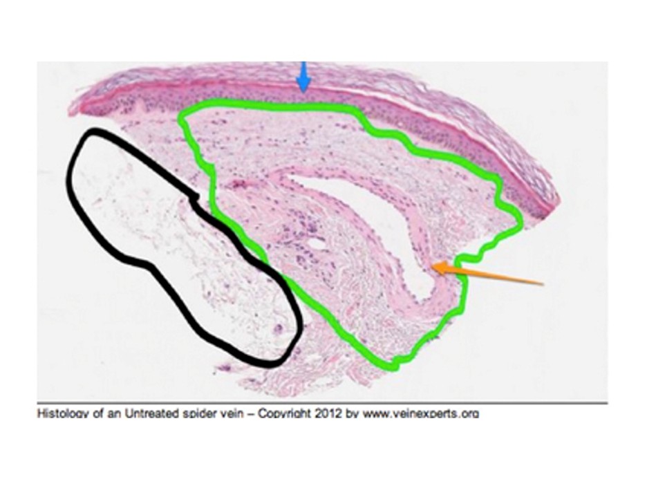
- Histology of a blue telangiectasia. Orange arrow points to telangiectasia. The area within the green outline is reticular dermis.
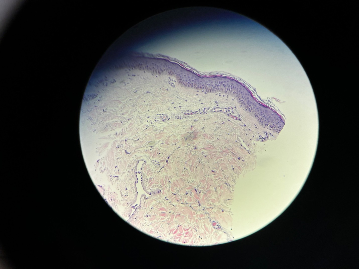
- Red telangiectasia in proximity of subpapillary plexus.
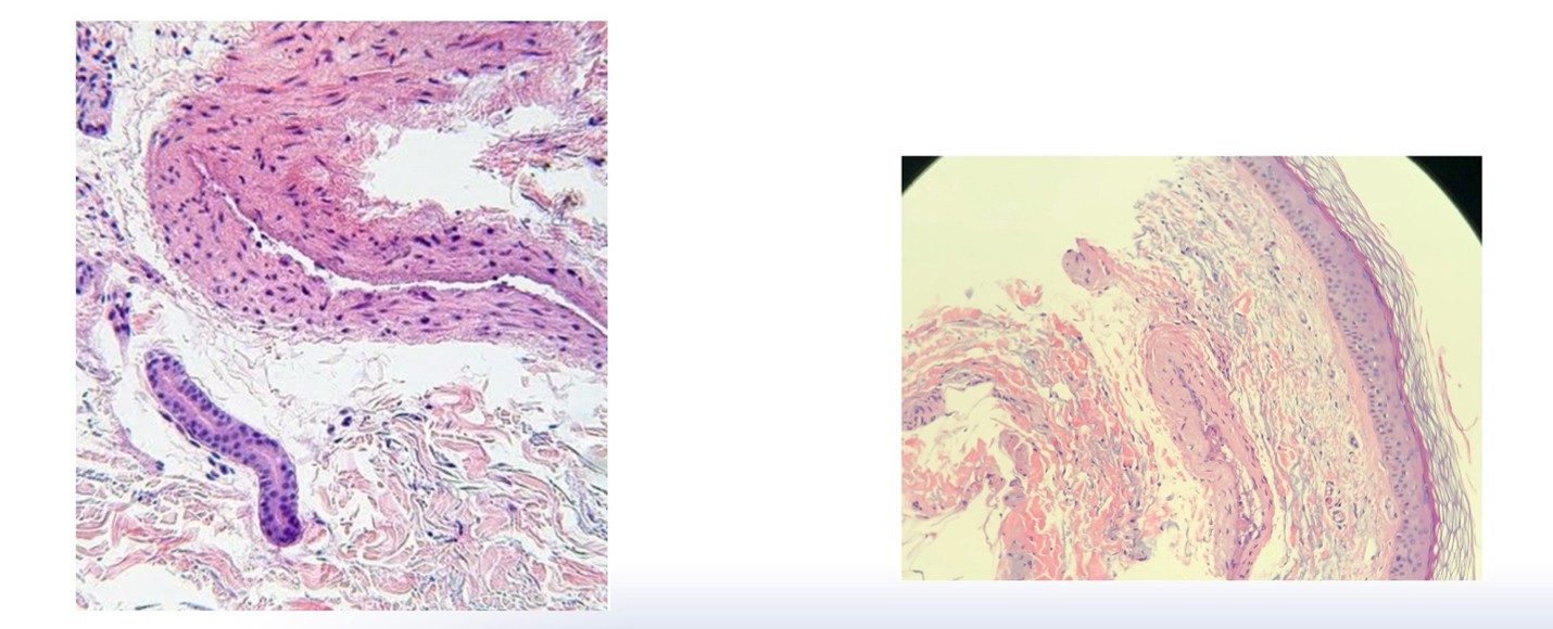
- Sotradecol® 0.1% diluted with normal saline on left. Sotradecol® 0.1% diluted with bacteriostatic water.
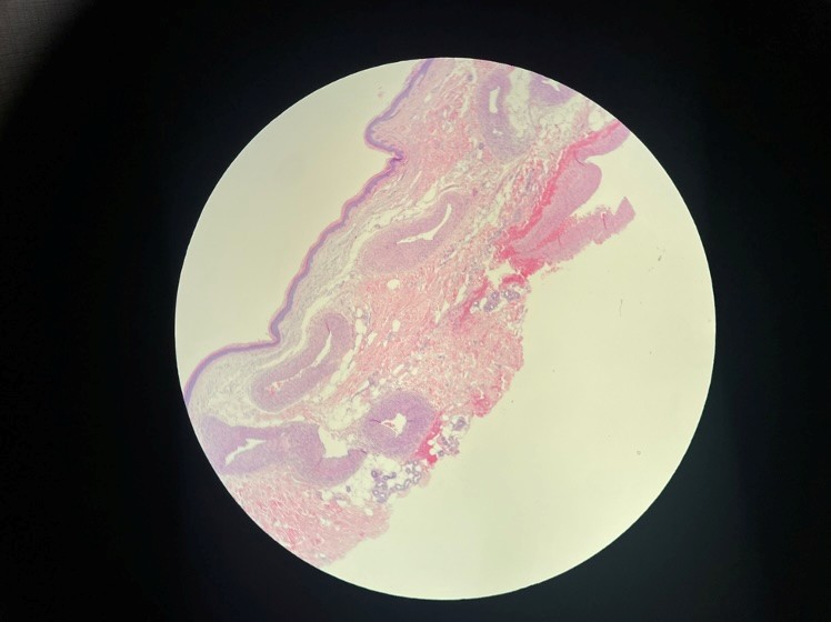
- Aneurysmal dilatation of spider veins.
What Type of LASER and How Much Energy Should Be Used

Rodrigo Kikuchi, MD, PhD, MBA
Angiologist and Vascular Surgeon
Choosing a laser for fine veins is not simply a matter of using more power. The real question is whether the wavelength, spot size, pulse duration, fluence, and cooling strategy are properly matched to the vessel being treated. And don’t forget the skin type in this equation…
Wavelength defines how light interacts with the target chromophore and how deeply energy can penetrate. Shorter wavelengths, such as KTP 532 nm and pulsed dye laser 585–595 nm, have strong hemoglobin absorption and are useful for superficial red vessels, but their penetration is limited. Alexandrite 755 nm offers intermediate penetration, while long-pulsed Nd 1064 nm penetrates more deeply and can reach subdermal and reticular veins, making it especially relevant for lower-limb telangiectasias and small reticular veins.¹
Energy selection should start with the vessel, not the machine. Very small dermal vessels may require smaller spots, shorter pulses, and higher fluence. Larger and deeper vessels usually require larger spots, longer pulses, and lower fluence to heat the vessel wall more uniformly. As a practical guide for 1064 nm Nd:YAG laser, dermal veins under 0.5 mm may require approximately 1–2 mm spot sizes, 5–10 ms pulses, and 250–300 J/cm². Telangiectasias between 0.5 and 1 mm may respond to 3–4 mm spot sizes, 10–15 ms, and 100–175 J/cm². Subdermal veins of 1–2 mm and reticular veins of 2–3 mm generally require longer pulses, larger spots, and lower fluence.
However, fixed settings are not enough. Histologic data show that even telangiectasias do not respond consistently to identical parameters.² The endpoint matters: vessel spasm, darkening, narrowing, or disappearance may be more useful than a rigid number on the screen. Cooling is essential to protect the epidermis, improve comfort, and allow effective vascular heating with a better safety margin.
The best setting is therefore not universal. It is individualized according to wavelength, vessel depth and diameter, skin type, device limitations, cooling, and the clinical endpoint. Anyone performing laser therapy for telangiectasias and reticular veins must understand how to balance the many variables involved in this complex therapeutic equation.
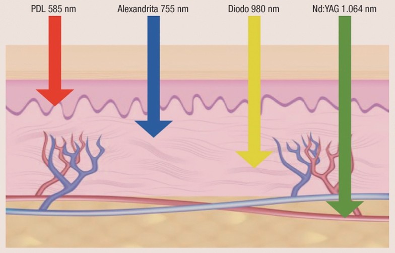
Figure 1. Schematic showing penetration depth of 532 nm, 595 nm, 755 nm, and 1064 nm lasers. Different wavelengths reach different vascular targets: superficial telangiectasias require a different strategy from deeper reticular veins. Source: Kikuchi R. Settings for vascular lesions with transcutaneous laser. In: Kikuchi R. Laser in Angiology and Vascular Surgery. Rio de Janeiro: DiLivros, 2024. p123-140
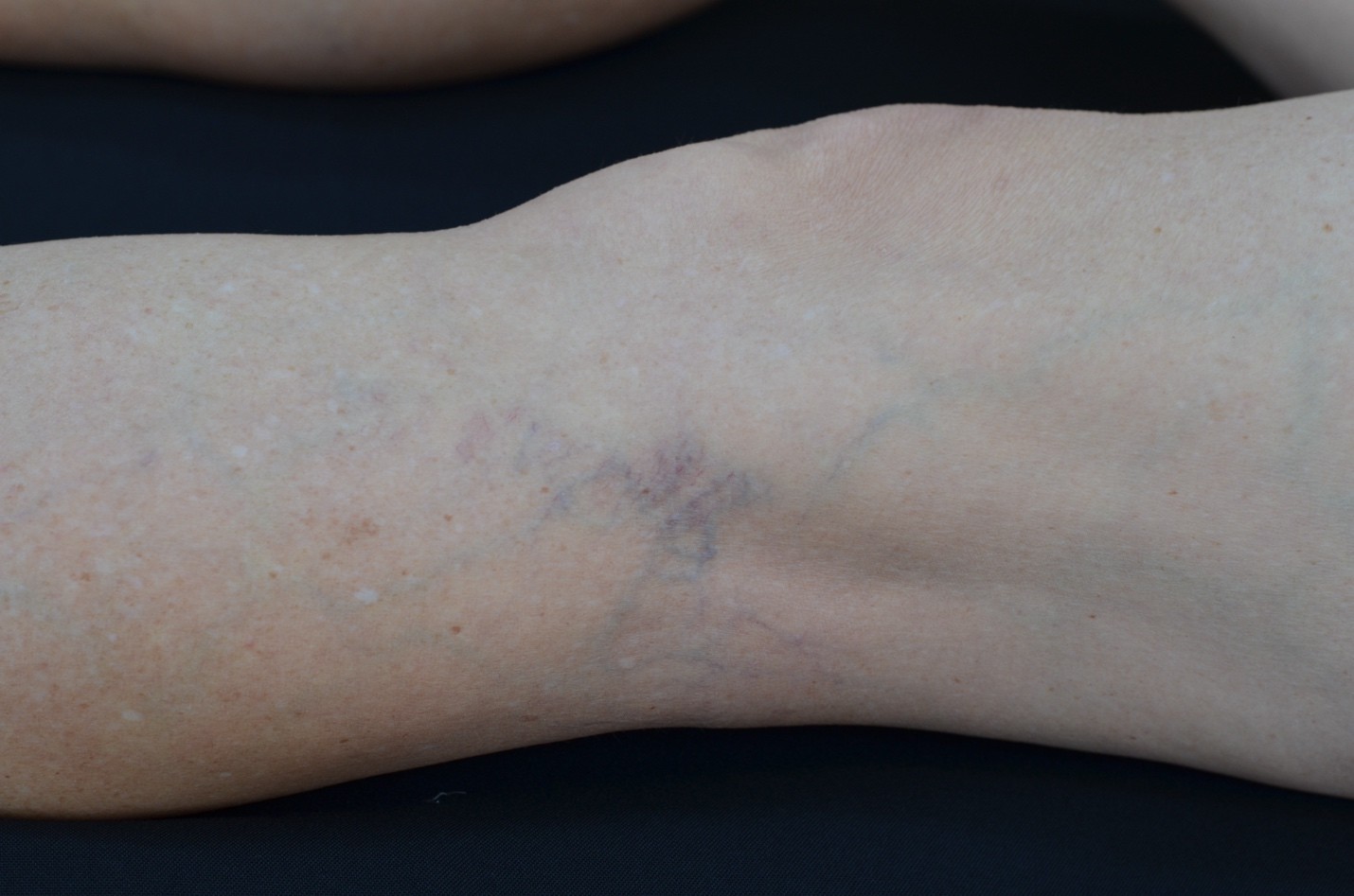
Figure 2. Clinical illustration of superficial telangiectasias and deeper reticular veins. Laser parameters should be selected according to vessel diameter, depth, and color.
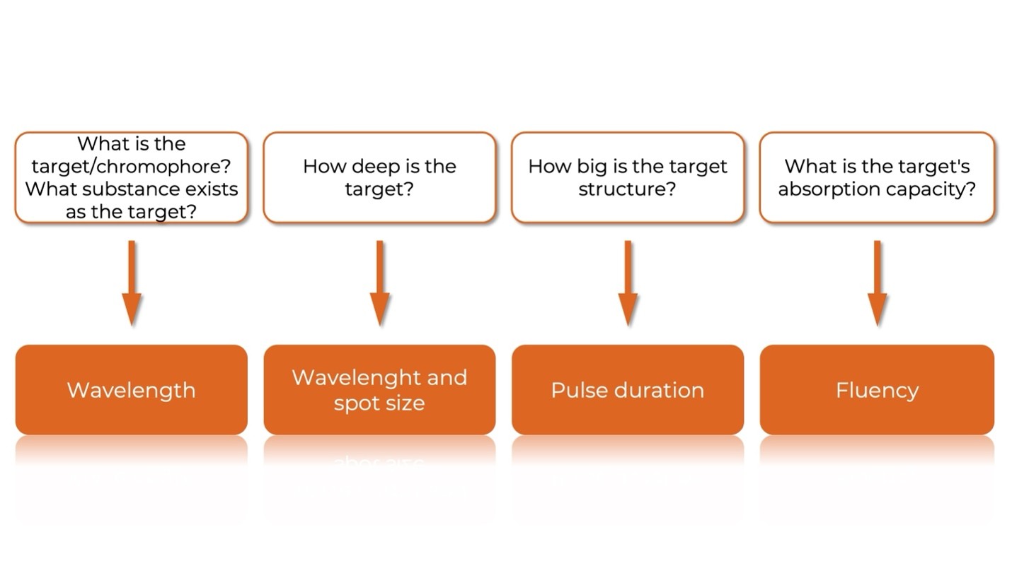
Figure 3. Rationale for selecting laser parameters in the treatment of veins with a 1064-nm Nd:YAG laser. Source: Kikuchi R. Settings for vascular lesions with transcutaneous laser. In: Kikuchi R. Laser in Angiology and Vascular Surgery. Rio de Janeiro: DiLivros, 2024. p123-140
References:
- Almeida JI, Raines JK. Laser ablation of cutaneous leg veins. Perspect Vasc Surg Endovasc Ther 2008;20(4):358-366
- Kikuchi R, Grill MH, Caffaro RA, et al. Effects of long-pulsed 1064-nm Nd laser on telangiectasias and reticular veins: a human in-vivo histological study. Int Angiol 2023;42(6):457-464
- Ianosi G, et al. Comparative study in leg telangiectasias treatment with Nd laser and sclerotherapy. Exp Ther Med2019;17(2):1106-1112
- Kikuchi R. Settings for vascular lesions with transcutaneous laser. In: Kikuchi R. Laser in Angiology and Vascular Surgery. Rio de Janeiro: DiLivros, 2024. pp123-140

Cosmetic Sclerotherapy: Tips and Tricks

Julia Motl, NP-BC
Stony Brook University Medical Center
Department of Vascular Surgery
Performing cosmetic sclerotherapy is a skill that takes dexterity, patience, and practice. It is not without its challenges. Telangiectasias can be difficult to treat, and telangiectatic matting is particularly challenging. Here are a few things I’ve learned over the years that may be helpful.
First, set expectations. It is our practice to tell patients that they can expect about an 80% improvement in the appearance of the treated veins. They must understand that the veins will not be gone, just less and less visible with each treatment. On average, we estimate that patients will require 3-6 sessions of sclerotherapy, with periodic sessions thereafter for maintenance.
Positioning is important for your comfort as well as the patient’s. Elevation of the legs on a wedge pillow will help promote closure of the vein networks and prevent thrombus formation post injection.
Bend your 30G needle to about a 45 degree angle with the bevel facing up. This will allow you a better angle of access into the vein while allowing you to rest your hand on the patient for stability. Always ensure you have the bevel of the needle facing up when you insert. Take note that sometimes the vessel may be so fine and so superficial that part of the bevel may be visible outside of the skin while you are injecting.
Play around with your lighting. Some practitioners like a direct spot light on the area. I find that I can visualize the vein better with indirect light, a bit off to the side of the patient.
Find an area within the telangiectasia where the vein looks straight and easy to enter. Remember you can milk the sclerosant into adjacent veins to fill them.
Pulling the skin taut helps gain entry into the vein.
As you are injecting, if you are not seeing the vein or the network of veins blanch, you’re not in. Stop and move to a new spot.
Slow injections will minimize matting.
Try to identify feeding reticular veins and treat those as well.
Large, dilated and dark purple telangiectasias benefit from foam sclerotherapy for better distribution and efficiency of the sclerosant while allowing for a lower total dose of sclerosant. I like to apply ice packs to these areas as well as any areas that are recalcitrant immediately post injection for a few minutes. Ice will also help alleviate some of the itching, burning discomfort from the injection.
It is very important to change your needle frequently. 30G needles can dull quickly, and dull needle is painful for the patient and will cause you difficulty getting into the vein.
Sclerotherapy can be fun and rewarding for both you and the patient. Good luck and have fun!
The 5 Biggest Mistakes Providers Make Before and After Sclerotherapy

Sclerotherapy is often viewed as a straightforward procedure, but achieving consistently excellent outcomes requires attention to details that extend well beyond the injection itself. While technique matters, some of the most common pitfalls occur before and after treatment.
- Failing to Perform a Thorough Venous Evaluation
Spider veins around the ankle warrant special attention, as they may indicate more advanced venous disease. In these patients, duplex ultrasound evaluation is often appropriate to identify underlying sources of venous hypertension. In addition, near-infrared vein visualization devices can help identify underlying feeder veins, perforators, and bifurcations that may not be readily visible on examination. The lateral reticular subdermal plexus is frequently involved in the development of spider veins. For optimal results, the lateral reticular perforator should be identified and treated, often with foam sclerotherapy, before addressing the visible telangiectasias. Failure to identify and treat the underlying source of reflux may lead to incomplete clearance and early recurrence.
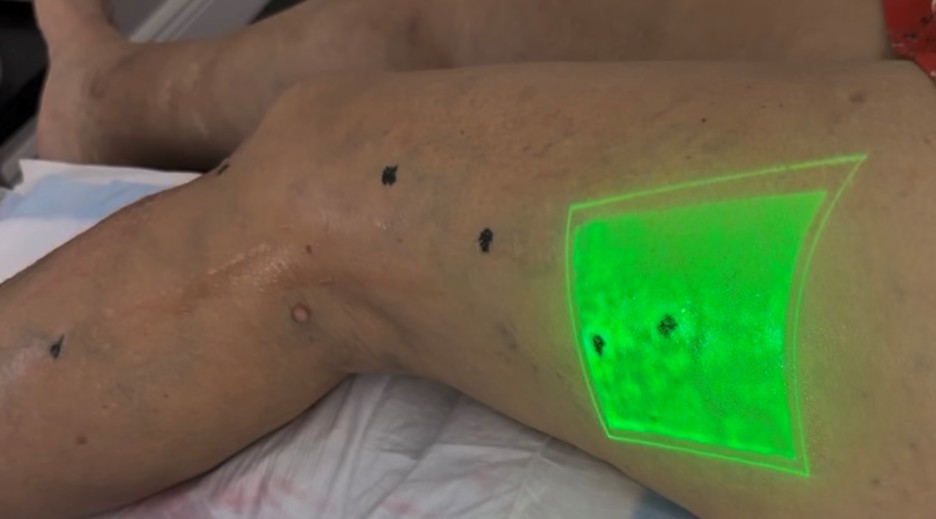
Figure 1. Careful pre-treatment evaluation, including visualization of feeder veins and perforators, helps improve treatment outcomes and reduce recurrence.
- Inadequate Patient Selection and Expectation Setting
Patients frequently expect complete clearance after a single session. Failing to discuss realistic timelines, the possibility of multiple treatments, and potential side effects such as bruising, hyperpigmentation, or matting can undermine patient satisfaction even when the procedure is technically successful.
- Neglecting Pre-procedure and Serial Photography
Baseline photographs are invaluable for documenting disease severity, monitoring progress, and demonstrating treatment success. However, photography should not be limited to the initial visit. Standardized photographs should be obtained at every treatment and follow-up visit using consistent lighting, positioning, and camera settings. Serial photography provides an objective record of improvement, helps guide treatment decisions, and allows providers to accurately assess outcomes over time.
- Providing Incomplete Post-treatment Instructions
Patients often remember only a fraction of what they hear during a visit. Clear written instructions regarding post-treatment care help improve adherence and reduce avoidable complications. Following treatment, application of a plant-based topical cream containing arnica may help reduce bruising, inflammation, and itching following vein treatments and procedures. Compression therapy should then be initiated as directed, along with recommendations for ambulation, activity restrictions, sun exposure, and follow-up care. Comprehensive post-treatment instructions can significantly improve patient satisfaction and clinical outcomes.
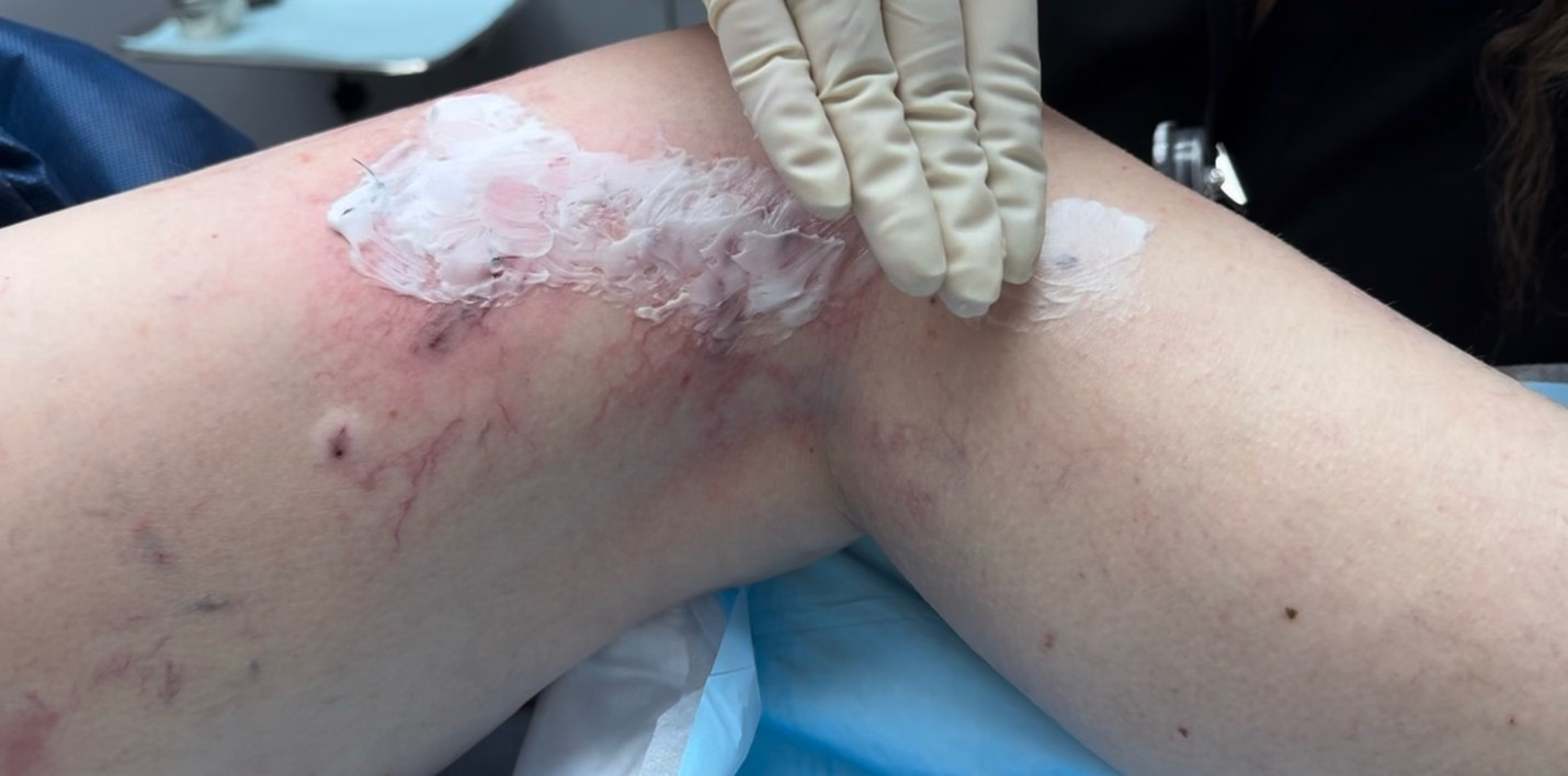
Figure 2. Clear post-treatment instructions, including topical therapy, compression, activity recommendations, and follow-up care, help minimize bruising and optimize patient outcomes.
- Failing to Schedule Appropriate Follow-up
Sclerotherapy should not be viewed as a one-time event. Follow-up visits allow providers to assess treatment response, identify trapped coagulum or adverse effects, determine the need for additional sessions, and reinforce patient expectations. Consistent follow-up often distinguishes excellent outcomes from merely acceptable ones.
Successful sclerotherapy begins with proper evaluation and continues long after the final injection. By focusing on patient selection, education, documentation, and follow-up, providers can improve outcomes, enhance patient satisfaction, and maximize the value of every treatment session.

