May/June Issue of vein specialist
Mixed Problems, Mixed Solutions
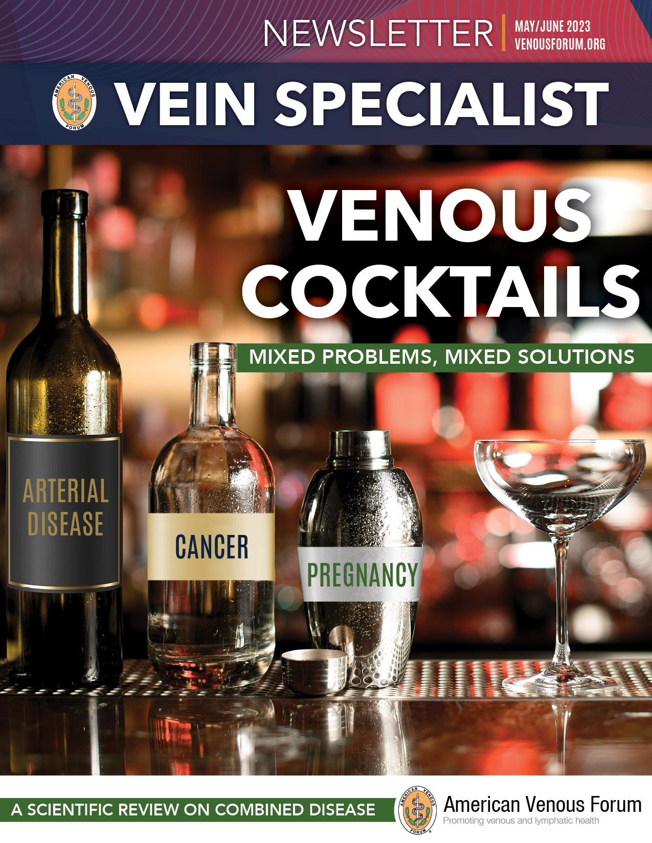
Table of Contents
Message from the Editor
Steve Elias, MD
Combined Disease
“Venous ulcers” and arterial disease
Robert Attaran, MD and Anil Hingorani, MD
Flavonoids for Treatment of Venous Ulcers
Monika Lecomte Gloviczki, MD, PhD
Venous Disease and Pregnancy: Navigating a Difficult Course
Qingwen Kawaji, MD and Maggie Arnold, MD, FACS
Venous Stasis Skin Change Mimickers
Eri Fukaya MD, PhD
The Queen is dead. Long live the King: How DOACs Replaced LMWHs in Cancer Thrombosis
Alfred Lee, MD, PhD
Leadership
President’s Message
Glenn Jacobowitz, MD
Meet Your 2023 American Venous Forum Board of Directors & Executive Committee
Allie Woodward
The Healthy Veins Book Launch Event in Canada
Beverly Chan, MD
Pediatric Lymphedema Champions
Margie Hopkins
Research Corner
Congratulations to the 2023 AVF Research Grant Winners!
Andrea Obi, MD
2024 AVF Research Grants – Application opening soon!
Membership
Copyright © 2023 by The American Venous Forum. All rights reserved.
EDITOR-IN-CHIEF
Steve Elias, MD
EXECUTIVE EDITOR
John Forbes, MBA
MANAGING EDITOR
Allie Woodward
PUBLICATION DESIGNER
Anthony Eaves
Message from the Editor
Steve Elias, MD
Editor-in-Chief, Vein Specialist
The Challenge? Nothing
Here’s the deal. This letter from the editor has no substance. Void of a story. But there is a challenge. I’ve got nothing to say. Like a Seinfeld episode. A show about nothing but maybe about everything. Why do we care about the inconsequential? So we don’t need to think about the real stuff. Minutiae becomes important for the moment. That’s where the danger lies or that’s where the creativity springs forth. Like Botticelli’s Birth of Venus (circa 1484-85; Uffize Gallery, Florence) emerging fully grown from the sea on the half shell. Here’s the challenge: Someone (one of you) write this letter. Here’s the schema:
- Identify 2 disparate concepts that may interest you.
- Elaborate on both separately.
- Do your research. Each concept will lead you down a rabbit hole. Jump in.
- Embrace this and cull one or two things from the rabbit hole.
- Go down another rabbit hole.
- Take a breath. Do whatever it takes to be creative: meditation, alcohol, yoga, weed, etc.
- Get out of your comfort zone. In life, to do anything new you need to feel comfortable with feeling uncomfortable.
- Once in the right frame of mind, meld the two disparate thoughts and the rabbit hole data.
- In between the two, discuss what’s in this issue of Vein Specialist.
- When done, read it to your family and friends.
- They may not like it. That’s their problem. Stick with it.
- Re-read your letter twice, out loud.
- Once satisfied, read it again.
- Finalize and send it to us. And don’t apologize. You’ve thought it through.
Now get writing and send it to me: [email protected]. The next letter is one of yours. No limits. No apologies. Just a challenge to be conquered. Do it. If you need help then you’re not up to the challenge. But if you do, have fun. It’s a letter about nothing. It’s a letter about everything.
I’m eager to hear what you have to say, but in the meantime, you’ll learn a lot from the authors who have provided valuable information in this issue about how to identify and treat patients with combined diseases, as well as meet AVF’s leadership.
“Venous ulcers” and arterial disease
Robert R. Attaran
Member
AVF Newsletter Committee

Anil Hingaroni
Member
AVF Newsletter Committee
Anil Hingorani
Member, AVF Newsletter Committee
Whilst symptom relief is a major goal for the venous practitioner, few endpoints are as important as venous ulcer healing. In a nutshell, we were taught that ulcers with an ischemic pattern required arterial revascularization.Ulcers within the gaiter zone were typically venous and required compression and correction of reflux and/or obstruction.
However, some patients, particularly the older age ones, have “mixed ulcers.” They develop ulceration around the ankle in the setting of concurrent peripheral arterial disease (PAD). There has been reticence to recommend compression for such patients. Nevertheless, in one survey of wound care practitioners, 80% would use compression for such mixed ulcers but only if the ankle brachial pressure index (ABI) was 0.8 or better (1). However, these mixed ulcers do take longer to heal (2).
There has been some reassuring data published on compression for venous ulcers in the setting of PAD. Mosti (2012) applied compression to 25 mixed ulcers patients. The patients had ankle pressures of ³60mmHg, toe pressures ³30mmHg and ABIs 0.5-0.8. Utilizing inelastic multi-later compression, perfusion to the peri-ulcer skin and foot was not compromised until the interface pressure was increased above 40 mmHg (3). In another small study, amongst patients without critical limb ischemia and ABI ranging from 0.5-0.8, inelastic compression at 30-40mmHg appeared to be tolerable and safe over 14 days (4). Mosti also reported that patients with mixed ulcers healed following only therapy for the venous pathology, plus compression (2).
Our approach to venous ulcer patients with concurrent PAD, then, was to treat the anatomic venous pathology (reflux or obstruction) and to apply compression. There was just one small problem: it didn’t work. At least, not as quickly as one had hoped. And when it did, recurrence rates were high. Often the ulcers persisted until arterial revascularization was carried out.
This observation is supported by Georgopoulos where amongst patients with mixed venous ulcers (0.5<AB<0.75) arterial revascularization was associated with faster ulcer healing. Eight weeks faster, in fact (16.6 v 24.7 weeks) (5). Similarly, Lantis observed faster ulcer healing with initial revascularization followed by compression (6).
None of the studies we’ve referenced were prospective control trials. Notwithstanding, a shift towards an “arterial revascularization first” approach appears to have worked better. Lastly, an additional consideration amongst such patients is to preserve the great saphenous vein in case it may be required for arterial bypass someday.
Patient with both arterial dna venous disease.
References:
- Woo KY, Sears K. Knowledge, Attitude, and Practice in the Management of Mixed Arteriovenous Leg Ulcers. Int J Low Extrem Wounds. 2016 Mar;15(1):52-7. doi: 10.1177/1534734615626626. Epub 2016 Jan 24. PMID: 26811376.
- Mosti G, Cavezzi A, Massimetti G, Partsch H. Recalcitrant Venous Leg Ulcers May Heal by Outpatient Treatment of Venous Disease Even in the Presence of Concomitant Arterial Occlusive Disease. Eur J Vasc Endovasc Surg. 2016 Sep;52(3):385-91. doi: 10.1016/j.ejvs.2016.06.004. Epub 2016 Jul 28. PMID: 27476154.
- Mosti G, Iabichella ML, Partsch H. Compression therapy in mixed ulcers increases venous output and arterial perfusion. J Vasc Surg. 2012 Jan;55(1):122-8. doi: 10.1016/j.jvs.2011.07.071.
- Ladwig A, Haase H, Bichel J, Schuren J, Jünger M. Compression therapy of leg ulcers with PAOD. Phlebology. 2014 May;29(1 suppl):7-12. doi: 10.1177/0268355514529507. Epub 2014 May 19. PMID: 24843079.
- Georgopoulos S, Kouvelos GN, Koutsoumpelis A, et al. The effect of revascularization procedures on healing of mixed arterial and venous leg ulcers. Int Angiol 2013;32(4):368-74.
- Lantis JC, 2nd, Boone D, Lee L, Mendes D, Benvenisty A, Todd G. The effect of percutaneous intervention on wound healing in patients with mixed arterial venous disease. Ann Vasc Surg 2011;25(1):79-86. DOI: 10.1016/j.avsg.2010.09.006.
Flavonoids for Treatment of Venous Ulcers

Monika Lecomte Gloviczki, MD, PhD
Research Fellow, Emeritus, Department of Internal Medicine
Gonda Vascular Center
Mayo Clinic
Rochester, MN

M. Mark Melin, MD, FACS, RPVI, FACCWS
Wound Healing Institute
M Health Fairview
Edina, MN

Emily Iker, MD
Lymphedema Center
Santa Monica, CA
John A. Chuback, MD, FACS, RPVI, RVT, RPhS
Chuback Vein Center
Paramus, NJ
Patients with chronic venous disease (CVD) and venous leg ulcers (VLUs) have a decreased quality of life. Olmsted County epidemiologic studies (1) demonstrated VLU prevalence (CEAP Class C5 and C6) did not decrease despite progress in minimally invasive endovenous technologies and adoption of early intervention principles for VLU patients (EVRA trial) (2). In a recent retrospective review of patients with VLUs, radiofrequency ablation (3) and concomitant ultrasound-guided foam sclerotherapy of perforating veins improved ulcer healing, though healing was only 56% at 6 months and recurrence rates were 23% after 3 years.
Flavonoids, prescribed worldwide for CVD, improve venous tone, venous wall protection, lymphatic drainage, hemmorhagic disorders, and demonstrate antioxidant and anti-inflammatory properties. This family of venoactive compounds includes diosmin, micronized purified flavonoid fraction (MPFF, 90% diosmin, 10% hesperidin), rutin, and rutosides. MPFF and hydroxyethylrutosides (HR) were studied as adjunctive therapy to compression and local wound care in VLU treatment.
The efficacy of MPFF was evaluated in a meta-analysis of five randomized controlled trials (RCTs) including data from 723 patients (4). At 6 months, ulcer healing was 61.3% in the MPFF group versus 47.7% in the control group (p=0.03). The chance of healing was 32% higher in patients treated with MPFF than in those managed by conventional therapy alone (RRR: 32%). Time to healing was 16 weeks in the MPFF group versus 21 weeks in the control group (p = 0.0034).
The efficacy of flavonoids in VLU patients was analyzed in a Cochrane review (5). The analysis included 5 RCTs with MPFF and 4 with HR, totaling 1075 participants. MPFF significantly improved the healing rate (RR 1.36). The issue of possible publication bias was raised, despite an independent meta-analysis that used the pulled electronic databases and favored the MPFF group. The three HR RCTs (279 patients) demonstrated a significant effect of HR on ulcer healing (RR 1.70). However, the reporting quality was judged as poor.
The flavonoid trial results for patients with VLUs demonstrate strong evidence for increased healing rates with MPFF as an adjunct to standard therapy. The value of HR for VLUs also appeared significant, but further trials are suggested. Adjuvant pharmacologic treatment, combined with conventional measures, may improve VLU closure rates and decrease ulcer recurrence.
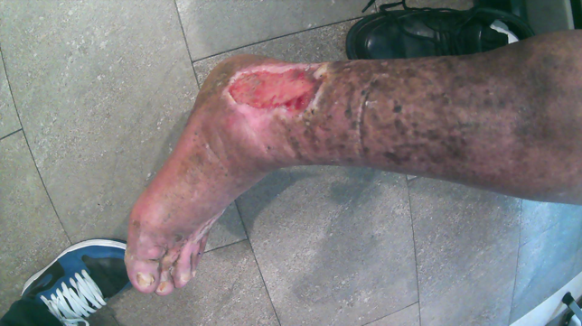
Clinical case (courtesy of Dr. Mark Melin):
A 74-year-old male presented with a chronic left VLU (Figure 1).
The past medical history included adult-onset diabetes, arterial hypertension, BMI of 40, no tobacco use, a normal ankle brachial index, and prior left great saphenous vein ablation. A venous competency study identified multi-level deep venous incompetency, ablated left saphenous, competent small saphenous and absent pathologic perforators. Incompetent varicosities were treated with foam sclerotherapy.
Compression therapy was fuzzy wale 8 mmHg compression under inelastic Velcro compression.
Wound care was pure hypochlorous acid soak for 10 minutes daily to decrease biofilm, with daily application of ovine foregut extracellular matrix.
Adjunctive micronutrients included Vitamins A, C, D, B12, B6, folate, zinc and MPFF (Vein Formula 1000), 500 mg twice daily.
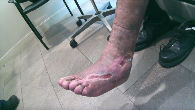
When healed (Figure 2), the patient was transitioned to compression stockings, ceramide-based pH 5.5 lotion, daily walking, and a bariatric medical consultation.
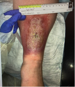
Figure 1. Before treatment, left leg, CEAP C6
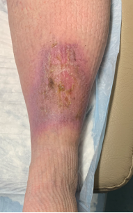
Figure 2. Three months after initiation of treatment, left leg, CEAP C5
References
- Gloviczki ML, Kalsi H, Gloviczki P, Gibson M, Cha S, Heit JA. Validity of International Classification of Diseases, Ninth Revision, clinical modification codes for estimating the prevalence of venous ulcer. J Vasc Surg Venous Lymphat Disord2014;2(4):362-7. DOI: 10.1016/j.jvsv.2014.03.002.
- Gohel MS, Heatley F, Liu X, et al. Early versus deferred endovenous ablation of superficial venous reflux in patients with venous ulceration: the EVRA RCT. Health Technol Assess2019;23(24):1-96. DOI: 10.3310/hta23240.
- Sermsathanasawadi N, Jieamprasertbun J, Pruekprasert K, et al. Factors that influence venous leg ulcer healing and recurrence rate after endovenous radiofrequency ablation of incompetent saphenous vein. J Vasc Surg Venous Lymphat Disord2020;8(3):452-457. DOI: 10.1016/j.jvsv.2019.11.003.
- Coleridge-Smith P, Lok C, Ramelet AA. Venous leg ulcer: a meta-analysis of adjunctive therapy with micronized purified flavonoid fraction. Eur J Vasc Endovasc Surg2005;30(2):198-208. DOI: 10.1016/j.ejvs.2005.04.017.
- Scallon C, Bell-Syer SE, Aziz Z. Flavonoids for treating venous leg ulcers. Cochrane Database Syst Rev2013(5):CD006477. DOI: 10.1002/14651858.CD006477.pub2.
