What is Vein Disease?
Find relevant information here.
Current Page →
What is Chronic Venous Disease?
Chronic venous disease (CVD) is a prevalent and multifaceted medical condition affecting the venous system, particularly the veins of the lower extremities. It encompasses a spectrum of venous disorders, ranging from mild cosmetic concerns to severe and debilitating conditions. CVD arises due to impaired blood flow within the venous system, leading to a variety of symptoms and potential complications. This page aims to provide a comprehensive overview of CVD, including its causes, risk factors, clinical manifestations, diagnostic approaches, and management strategies.
Anatomy and Physiology of the Venous System
Knowledge of the anatomy and physiology of the venous system is essential to understanding chronic venous disease. Veins are blood vessels responsible for transporting deoxygenated blood back to the heart. In the lower extremities, veins play a vital role in overcoming gravity, aided by one-way valves that prevent backflow. The calf muscles acting as a pump assist in propelling venous blood toward the heart during muscle contraction. Inadequate venous valve function or impaired muscle pumping can lead to venous stasis (pooling of blood in the leg) and, subsequently, chronic venous disease. Weak or damaged valves within the veins result in retrograde blood flow (toward the feet instead of back up to the heart), which is called venous reflux, and leads to venous hypertension (“high pressure” in the legs) and dilation of veins.
Causes and Risk Factors
Numerous factors contribute to the development of chronic venous disease. Some of the primary causes and risk factors include:
- Genetics: A family history of venous disorders can increase an individual’s susceptibility to CVD.
- Age: The risk of CVD increases with age due to natural wear and tear on the venous system.
- Prolonged sitting or Standing: Occupations that involve extended periods of sitting or standing can impede venous blood flow, contributing to venous insufficiency.
- Obesity: Excessive body weight puts additional pressure on the veins and can impair venous return.
- Pregnancy: Hormonal changes during pregnancy and the pressure of the growing uterus on pelvic veins can lead to venous insufficiency.
- History of deep vein thrombosis (DVT): A past episode of DVT can damage valves and veins, increasing the risk of developing CVD.
Clinical Manifestations
The clinical presentation of chronic venous disease can vary widely, ranging from no symptoms to severe symptoms. Common signs and symptoms include:
- Varicose veins: Enlarged, twisted, and bulging veins that are often blue or purple, commonly seen in the legs.
- Leg heaviness and fatigue: Patients may experience a sensation of heaviness or tiredness in the legs, especially after prolonged standing or sitting.
- Edema: Swelling in the lower legs and ankles due to fluid accumulation caused by venous congestion.
- Skin changes: Chronic venous disease can lead to skin problems such as venous eczema or dermatitis (reddish skin discoloration, itching, and dryness).
- Hemosiderin staining: Brownish discoloration of the skin in the calf.
- Lipodermatosclerosis: A condition characterized by fibrosis and hardening of the skin and subcutaneous tissues.
- Venous ulcers: In advanced stages, open wounds may develop in the calf/ankle.
- Other complications (Chapter 7, see also Phlebitis and Bleeding on the website):
- Bleeding: Veins very near the skin surface can tear and bleed after scratching or minor trauma.
- Phlebitis: Inflammation of a superficial vein (varicose vein) with the formation of a blood clot.
Diagnostic Approaches
Accurate diagnosis is essential for appropriate management. Physicians use various diagnostic tools to assess chronic venous disease:
History taking: Providers will ask questions such as when veins first appeared; symptoms and activities that make them worse or better; how symptoms affect one’s job and/or activities of daily living; and about compression stocking use. They will also review personal and family history of varicose veins; blood clots and clotting disorders; history of leg trauma and/or orthopedic history; any conditions that affect leg/ankle/foot movement; and pregnancy history.
Physical examination: A thorough clinical examination can reveal visible signs of varicose and spider veins, skin changes, swelling.
Duplex utrasound (most common): This non-invasive imaging technique allows visualization of blood flow in the veins and helps identify weak or damaged valves, abnormal blood flow, and blood clots.
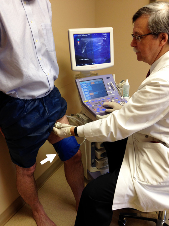
To examine the function of the vein valves, the ultrasound exam is done in the standing position. An inflation cuff (arrow) is often used to quickly push the blood flow upward in order to measure how rapidly the vein valves close (Photo courtesy of John Blebea, MD) (The Healthy Veins Book, 2023, p. 41, fig. 8-6).
Management Strategies
The management of chronic venous disease aims to relieve symptoms, prevent complications, and improve patients’ quality of life. The treatment options include:
- Lifestyle modifications: Encouraging regular physical activity, weight management, and avoiding prolonged periods of sitting or standing.
- Compression therapy: Wearing compression stockings can improve venous blood flow, reduce edema, and alleviate symptoms.
- Sclerotherapy: Injection of a sclerosant solution into varicose veins to cause their closure and subsequent reabsorption.
- Phlebectomy: Removal of bulging varicose veins, which can be performed in the office.
- Surgical Interventions: For severe cases or complications, surgical procedures like vein ligation and stripping may be considered.
- Endovenous ablation: Minimally invasive procedures, such as laser or radiofrequency ablation, mechano-chemical ablation, and cyanoacrylate glue sealant, are used to treat underlying venous reflux.
Chronic venous disease is a prevalent condition with a wide range of clinical presentations and potential complications. Early recognition, appropriate diagnosis, and timely intervention are crucial in preventing disease progression and improving outcomes. By implementing a combination of lifestyle modifications, conservative measures, and medical interventions, your physician or non-physician provider can help you effectively manage CVD and enhance your quality of life.
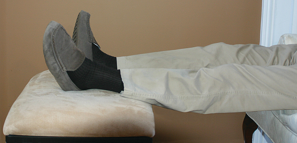
Keeping the legs elevated on a footstool while sitting will help control swelling and fluid buildup in the legs (Photo courtesy of John Blebea, MD) (The Healthy Veins Book, 2023, p. 46, fig. 9-2).
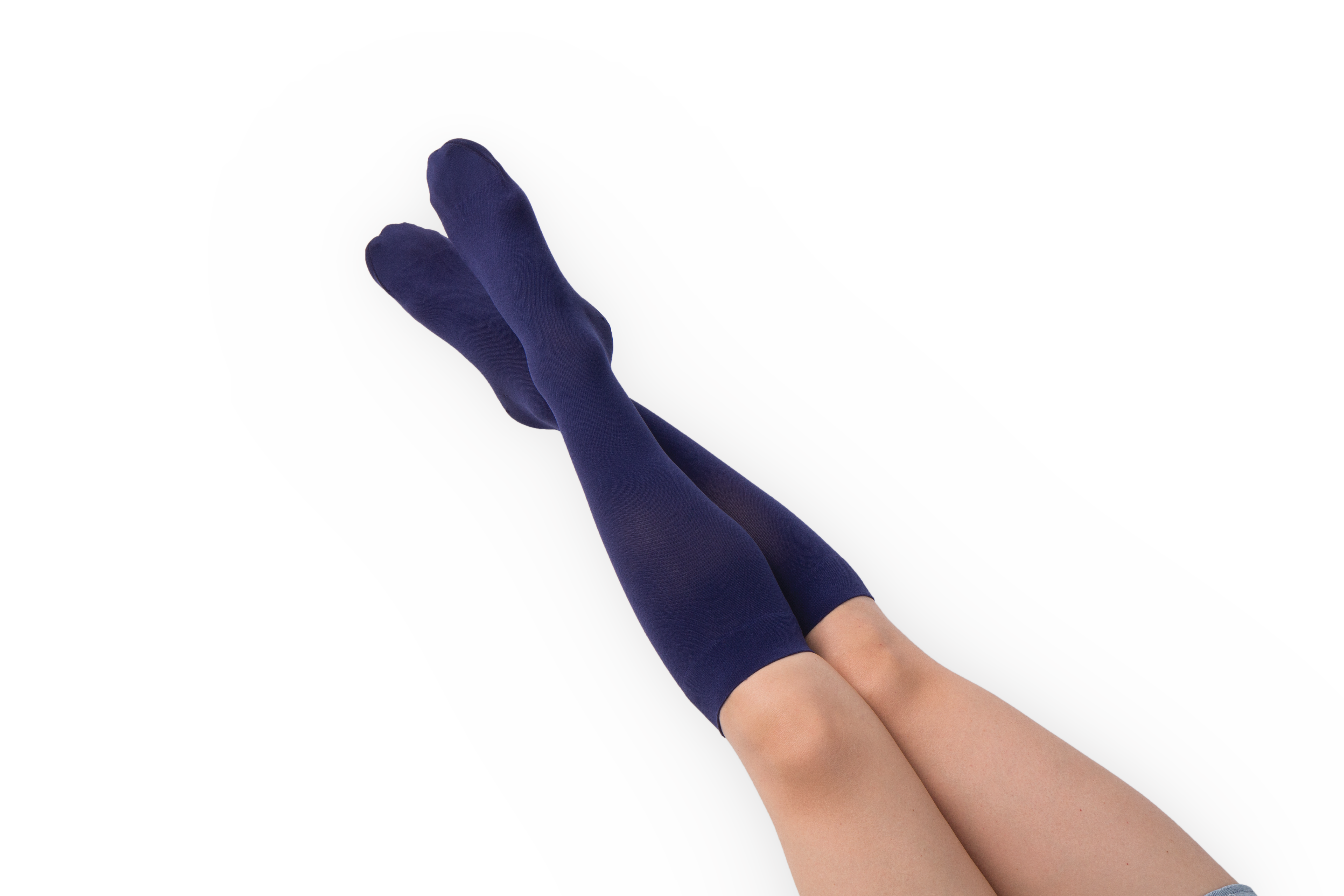
Compression stockings, particularly those to the knee, are available in multiple color and fabric options (The Healthy Veins Book, 2023, p. 47, fig. 9-4).
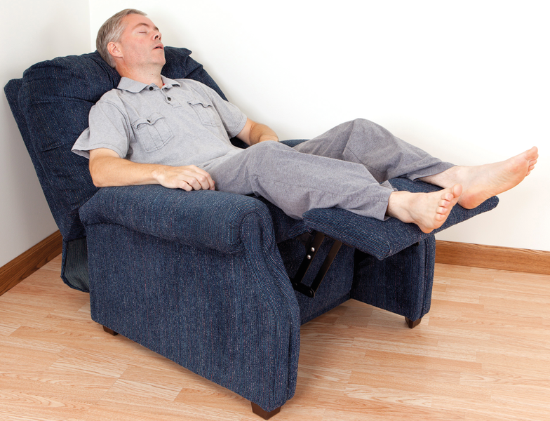
When at home and neither sleeping nor walking, sitting in a recliner chair can provide a lot of elevation for the legs to keep the swelling controlled (The Healthy Veins Book, 2023, p. 135, fig. 27-2).
Chapter 4: Varicose Veins
Chapter 5: Who Gets Varicose Veins?
Chapter 6: Symptoms of Varicose Veins and Bad Vein Valves
Chapter 9: Healthy Habits
Chapter 11: Compression Stockings and Wrappings
Chapter 15: Radiofrequency Ablation
Chapter 16: Laser Ablation
Chapter 17: Mechano-chemical Ablation of Veins
Chapter 18: Sealing the Veins with Glue
