

Vein Specialist
2026September/October
The New Frontier: Expanding Horizons of PE Management in 2026

Evolution in the Management of Complex and Advanced Presentations of Acute Pulmonary Embolism: Clot-in-Transit and VA-ECMO

Charles B. Ross, MD, FACS, DFSVS
Vascular/Endovascular Surgery
Pulmonary Embolism Response Team
Piedmont Atlanta Hospital, Atlanta, GA

Andrew J.P. Klein, MD, FACC, FSCAI
Interventional Cardiology
Pulmonary Embolism Response Team
Piedmont Atlanta Hospital, Atlanta, GA

Christian Bermudez, MD, FACS
Cardiac and Thoracic Surgery
Surgical Director of Lung Transplant and CTEPH Program
Pulmonary Embolism Response Team
Piedmont Atlanta Hospital, Atlanta, GA
Management of acute pulmonary embolism (PE) has undergone momentous change over the past two decades. Recent publication of the 2026 ACC/AHA Guidelines for Management of Acute PE represent the most comprehensive approaches to care from diagnosis to post discharge status presently available.1 Much of the change in care of acute PE has been driven by new and improved interventional procedures aimed toward reduction of pulmonary artery obstruction to relieve the failing right ventricle (RV) guided by pulmonary embolism response team (PERT) collaboration. Two situations where percutaneous intervention and cardiac surgery intersect, clot-in-transit (CIT) and rescue utilization of VA-ECMO, have been part of the advancing evolution of care.
Clot-In-Transit
Acute thrombus identified within the superior or inferior vena cava and/or right atrium, originating from either the lower or upper extremities, may be technically considered CIT; but, the most frequent and dramatic scenario encountered involves a mobile, tumbling thrombus identified in the right atrium with associated acute PE, representing a thrombus load that has not yet embolized into the RV outflow tract but is at risk for doing so. In the landmark iCOOPER registry, a CIT was noted in 4.0% of patients, which is consistent with the more recent real-world registry of 12,000 patients from the PERT ConsortiumTM database, wherein the incidence was 4.9%.3 PE outcomes may be negatively affected by CIT, especially when a patent foramen ovale (PFO) is involved and thrombus extends into the left atrium. CIT occurs frequently enough that every PERT should expect to encounter such cases.
CIT may be identified on a presentation CTPA and/or transthoracic echo and is more common in patients with pre-existing congestive heart failure and in cases of severe to catastrophic PE. Although CIT was once considered an absolute indication for surgical embolectomy, our PERT management has evolved to the use of large-bore aspiration thrombectomy (LBAT) with TTE or intracardiac echo guidance in awake patients and TTE and/or TEE guidance in those already intubated. CIT in the setting of a PFO, especially with extension of thrombus into the left atrium, requires more scrutiny. Thrombus size, anatomical PFO considerations, and patient characteristics play an important role in the decision making. Although successful percutaneous management by LBAT plus cerebral protection has been reported, 4 surgical embolectomy remains the gold-standard for management. In all cases of CIT, cardiac surgical input to the PERT decision is essential. The 2026 AHA/ACC guidelines state that utilization of advanced therapies over anticoagulation alone is reasonable to reduce the risk of clinical deterioration in patients with acute PE, categories C3-E2, who have a free-floating right atrial thrombus or CIT (2a recommendation).1 The complexity of management as well as the immediate need for rescue VA-ECMO should a massive CIT suddenly embolize in the RV outflow tract, in our opinion, mandates management of patients with CIT to centers with open cardiac surgery and VA-ECMO capability.
Veno arterial – extracorporeal membrane oxygenation (VA-ECMO)
Most acute PEs, when anticoagulated, are survivable. The technologic advance that has had perhaps the greatest impact in salvaging otherwise fatal acute PEs has been VA-ECMO. Survival rates have been reported as high as 61% when initiated during CPR for acute PE.5 When all indications for VA-ECMO in PE including eCPR, pre-emptive bridging combined with advanced therapy, and VA-ECMO plus simple anticoagulation alone are considered, survival ranges from 60 to 70%.6 A remarkable survival rate of 96 % was reported in 2018 from the University of Maryland where i a VA-ECMO-first protocol for massive PE was incorporated. VA-ECMO currently carries a 2a recommendation for use in patients with Class E2 acute PE. The role of pre-emptive VA-ECMO for category D and E1 PE is patient/institution specific and unknown. VA-ECMO, which can be instituted remotely, is a powerful tool which comes with its own potentially fatal complications; therefore, its use seems best when applied through collaborative PERT care or pre-defined institutional protocols for patient selection.
In summary, advances such as LBAT for CIT and the role of VA-ECMO in cases of class D and E PE hold promise for continued reductions in morbidity and mortality from complex and life-threatening acute PEs and argue strongly for collaborative PERT management.
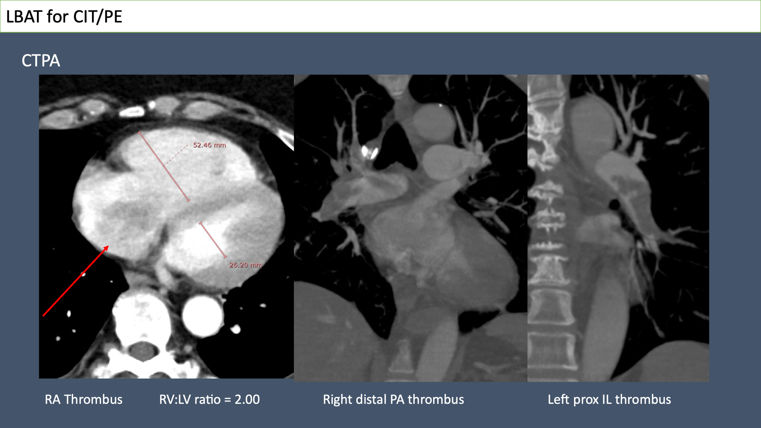
Figure 1: 62-year-old woman presented with category D1R acute PE. The initial CTPA suggested CIT with saddle PE and right ventricular strain.
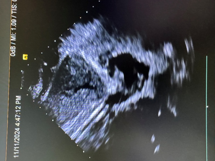
Figure 2a: Echo showing 24F Flowtriever ®(T24) approaching the RA thrombus.
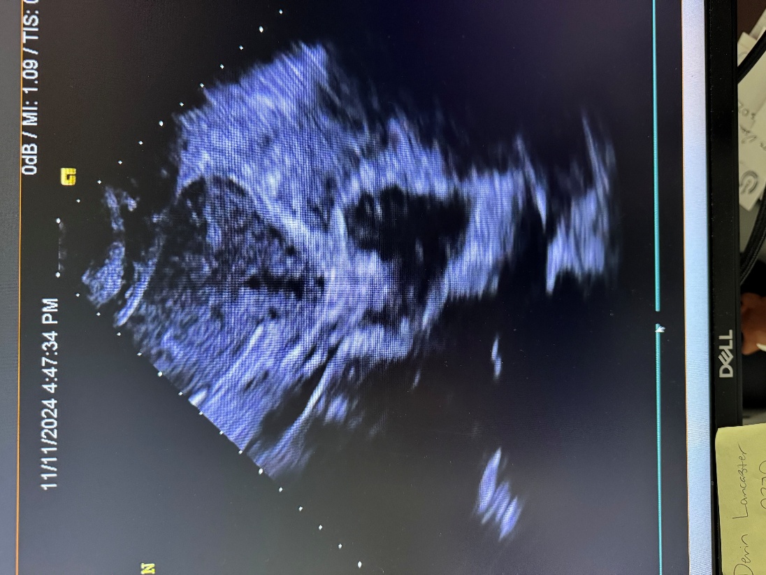
Figure 2B: Capture of the thrombus.
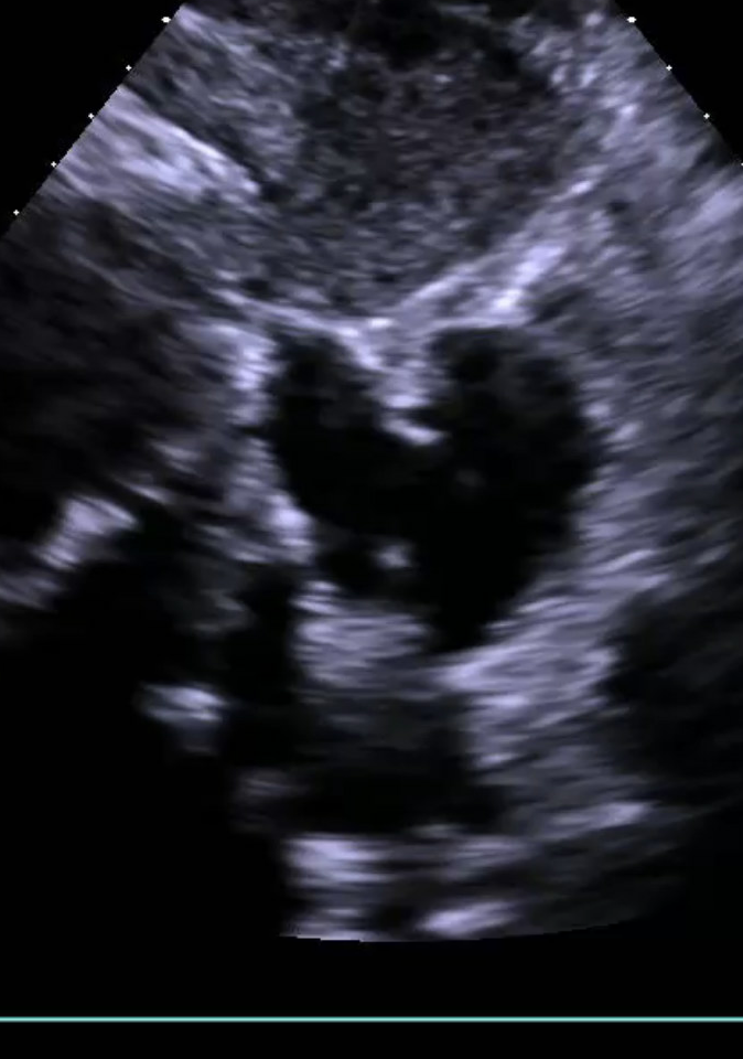
Figure 2C: Thrombus successfully removed after two pulls via the T24.
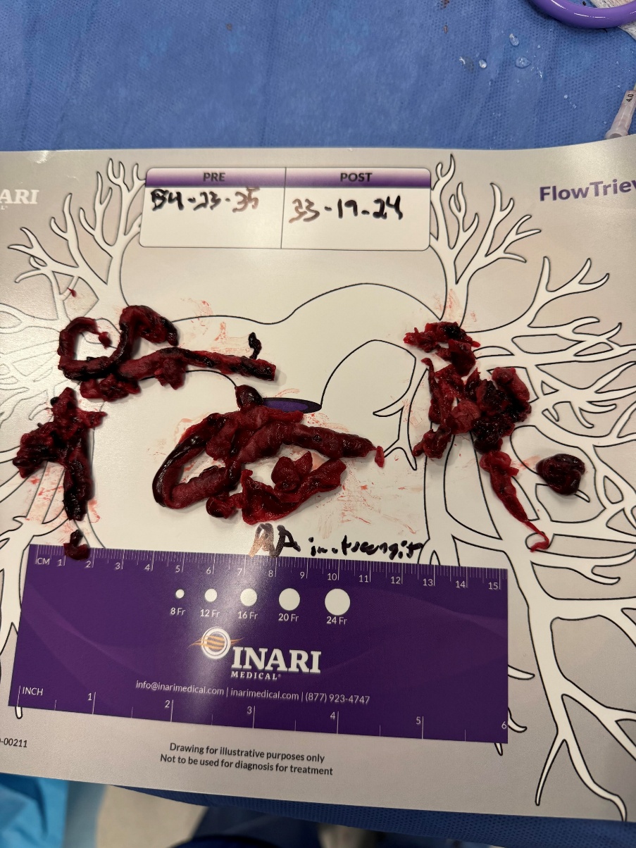
Figure 3: Specimen of thrombus successfully removed by LBAT procedure.
References
1. Creager MA, Barnes GD, Giri J, et al. 2026 AHA/ACC/ACCP/ACEP/CHEST/SCAI/SHM/SIR/SVM/SVN Guideline for the Evaluation and Management of Acute Pulmonary Embolism in Adults: A Report of the American College of Cardiology/American Heart Association Joint Committee on Clinical Practice Guidelines. Circulation 2026 Mar 24;153(12):e977-e1051. doi: 10.1161/CIR.0000000000001415. Epub 2026 Feb 19. Erratum in: Circulation 2026 Jul 14;154(2):e24. doi: 10.1161/CIR.0000000000001462. PMID: 41712677.
2. Torbicki A, Galié N, Covezzoli A, Rossi E, De Rosa M, Goldhaber SZ; ICOPER Study Group. Right heart thrombi in pulmonary embolism: results from the International Cooperative Pulmonary Embolism Registry. J Am Coll Cardiol. 2003 Jun 18;41(12):2245-51. doi: 10.1016/s0735-1097(03)00479-0. PMID: 12821255.
3. Keeling WB, Horbal S, Rosenfield K, et al. Pulmonary Embolism in the United States: A Report From the National PERT Consortium. J Soc Cardiovasc Angiogr Interv. 2026 Jun 2;5(7):105389. doi: 10.1016/j.jscai.2026.105389. PMID: 42502334; PMCID: PMC13400114.
4. Joseph S, Rengifo-Moreno P. Percutaneous Thrombectomy of Clot in Transit Lodged in the Patent Foramen Ovale Using the FlowTriever Device: Case Report. J Soc Cardiovasc Angiogr Interv. 2025 Aug 28;4(10Part A):103879. doi: 10.1016/j.jscai.2025.103879. PMID: 41268091; PMCID: PMC12629719.
5. Rali P, Moores L, Criner G, et al. Venoarterial Extracorporeal Membrane Oxygenation in Massive Pulmonary Embolism-Related Cardiac Arrest: A Systematic Review Critical Care Medicine. 2021;49(5):760-769. doi:10.1097/CCM.0000000000004828
6. Abdulaziz S MBBS, Kakar V, Kumar PG, et al. Mechanical Circulatory Support for Massive Pulmonary Embolism. J Am Heart Assoc. 2025 Jan 7;14(1):e036101. doi: 10.1161/JAHA.124.036101. Epub 2024 Dec 24. PMID: 39719427; PMCID: PMC12054433.
7. Pasrija C, Shah A, George P, et al. Triage and optimization: a new paradigm in the treatment of massive pulmonary embolism. J Thorac Cardiovasc Surg. 2018;156:672–681.

Sequelae of PE and PE Interventions

Vic Tapson, MD, FCCP, FRCP
Professor of Medicine
Division of Pulmonary and Critical Care
Cedars-Sinai Medical Center
Sr. Director, Medical Affairs
Stryker PV
Acute pulmonary embolism (PE) causes tremendous early mortality with approximately 100,000 deaths per year in the United States–amounting to one death every 5 minutes. In fact, several sources suggest that mortality may, in fact, be increasing over the past several decades.1,2
But potential longer-term sequelae cannot be ignored. Disagreements have arisen regarding terminology, and “post-PE syndrome” has often served as an umbrella term that includes symptoms due to deconditioning, anxiety, coexisting illnesses, and quality of life that persist for longer than 3 months after the initial acute PE event, as well as those that are derived directly from obstruction by chronic thromboembolism.3
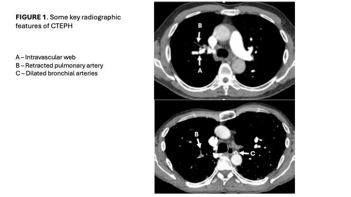
The most devastating form of post-PE syndrome is chronic thromboembolic pulmonary hypertension (CTEPH), which occurs in approximately 3% of acute PE patients. The time delay between onset of symptoms and diagnosis and referral to an expert pulmonary hypertension center is often more than one year, and the majority of patients present with advanced symptoms of dyspnea with exertion. While prognosis is poor in untreated patients, CTEPH can be cured by pulmonary endarterectomy, although only a handful of expert surgical centers in the US perform more than 50 of these cases per year. In certain Western European countries, there is often a single national expert center. In patients who are not candidates for surgical endarterectomy (often due to more distal obstruction), extensive experience now exists with balloon pulmonary angioplasty.4 A computed tomographic angiogram image showing key features of CTEPH is shown in Figure 1.
Persistent symptoms after acute PE should be evaluated with echocardiography as a screen to determine whether pulmonary hypertension (CTEPH) is present; if so, a more detailed evaluation including right-heart catheterization is undertaken. If there is no pulmonary hypertension, but persistent CT scan abnormalities are present, further evaluation is necessary to sort out the potential cause to determine whether the persistent vascular changes are, in fact, causing symptoms. Cardiopulmonary exercise testing has become increasingly useful in this regard.
Finally, we must determine if PE interventions such as catheter-directed thrombolysis or catheter embolectomy cause problematic sequelae, Large randomized clinical trials suggest that while complications can occur, including major bleeding or pulmonary artery perforation, these are very rare, and these procedures can avoid potentially catastrophic complications such as intracranial hemorrhage caused much more frequently by systemic thrombolysis.5
References:
1. Casey SD, Stubblefield WB, Luijten D, et al. Addressing the rising trend of high-risk pulmonary embolism mortality: Clinical and research priorities. Acad Emerg Med. 2024; 31:288-292.
2. Martin KA, Molsberry R, Cuttica MJ, et al. Time trends in pulmonary embolism mortality rates in the United States, 1999 to 2018. J Am Heart Assoc. 2020; 9(17) e016784.
3. Morris TA, Dakaeva K, Alotabi M, et al. Chronic thromboembolism phenotypes after acute pulmonary embolism: identification with the SEARCH algorithm. Eur Respir J 2026 68(2): 2501562.
4. Jais X, Tapson V, Fernandes TM, et al. Diagnosis of chronic thromboembolic pulmonary hypertension. J Heart Lung Transplant 2025; 44(7S): S1-S7.
5. Jaber WA, Gonsalves CF, Stortecky S, et al. Large-bore mechanical thrombectomy versus catheter-directed thrombolysis in the management of intermediate-risk pulmonary embolism: primary results of the PEERLESS randomized controlled trial. Circulation 2025; 151(5): 260-273.

Help advance treatment for chronic pelvic pain.
The EMBOLIZE Study, led by the SIR Foundation, in collaboration with the VIVA Foundation and Penumbra, is a first-of-its-kind randomized controlled trial studying ovarian and pelvic vein embolization for women with chronic pelvic pain due to pelvic venous disease.
This study aims to generate the high-quality evidence needed to:
Improve diagnosis and care for pelvic venous disease
Evaluate the impact of embolization on pain and quality of life
Expand awareness and referral pathways for patients suffering from chronic pelvic pain
We are currently seeking patients to participate in this important research effort.
By referring your patients to the study, you will help address a major unmet need and improve outcomes for millions of women suffering from this often overlooked and under treated condition.
Visit embolizetrial.com to learn more.
Biomarkers to Predict Thromboembolic Pulmonary Hypertension and Refine Patient Selection for Intervention

Elizabeth Andraska, MD, MSc
Assistant Professor of Vascular Surgery, University of Pittsburgh
Chronic thromboembolic pulmonary hypertension (CTEPH) is a rare but debilitating complication following acute pulmonary embolism (PE). CTEPH is characterized by persistent functional impairments and resting pulmonary hypertension following PE. The condition is associated with significant morbidity and impaired quality of life, positioning early risk stratification and diagnosis as a central objective for those studying CTEPH. While current biomarkers such as pro-BNP, troponin, and lactate help to stratify the acute PE patient into severity categories and identify those at risk for acute clinical decompensation, they are not as helpful at identifying those who will develop chronic complications of PE. Further, a biomarker that predicts CTEPH may help with clinical decision making to help determine which patients should be treated more aggressively with advanced catheter-based therapies.
Mechanistically, CTEPH is thought to arise from the failure of thrombus to completely resolve, which results in localized inflammation and fibrotic remodeling of the pulmonary arteries, chronic right heart strain, and abnormal microvascular overgrowth, ultimately resulting in higher arterial resistance. Collectively, these processes increase pulmonary vascular resistance and drive the transition from acute thromboembolic disease to a chronic obstructive vasculopathy.
Efforts to translate this pathophysiology into clinically applicable biomarkers have started to focus on mediators of cardiopulmonary inflammation and the thromboinflammatory axis. Although several biomarkers demonstrate statistically significant associations with CTEPH, none have been shown to function as a stand-alone diagnostic marker, and none have been used to guide interventional decision making. Given the diagnostic and mechanistic complexity of CTEPH, it is unlikely that a single biomarker will be sufficient to reliably predict disease progression. Rather, current findings may serve as a foundation for a more multimodal, precision- medicine-based approach to diagnosis of CTEPH following PE. If we are ultimately able to use biomarkers to identify patients at most risk for CTEPH, we may be able to more confidently and aggressively treat those patients with advanced catheter-based therapies to improve long term outcomes.
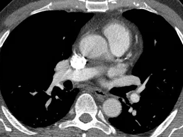
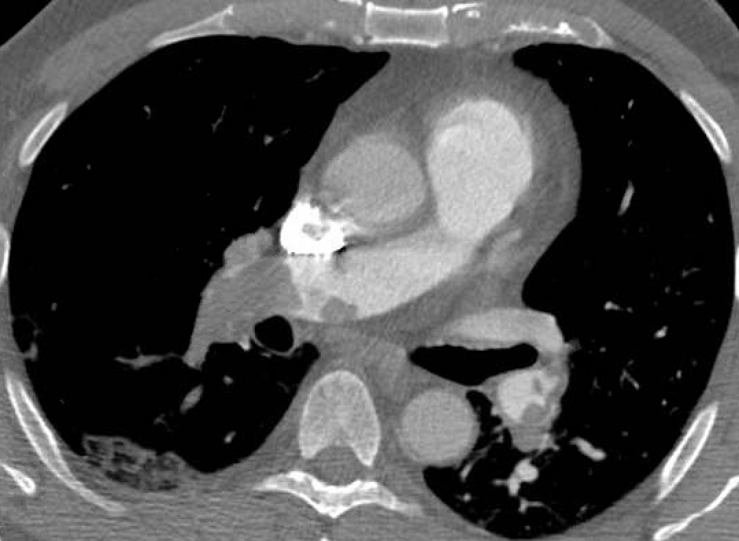
Figures 1 & 2: Improved PE thrombus burden after acute mechanical thrombectomy.
