
 Steve Elias, MD
 Ronald G. Bush, MD, FACS
 Test Testing, MD
 Test Testing, MD
Reach out! [email protected].
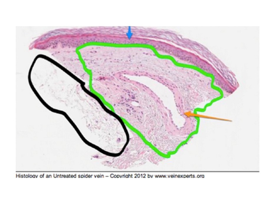
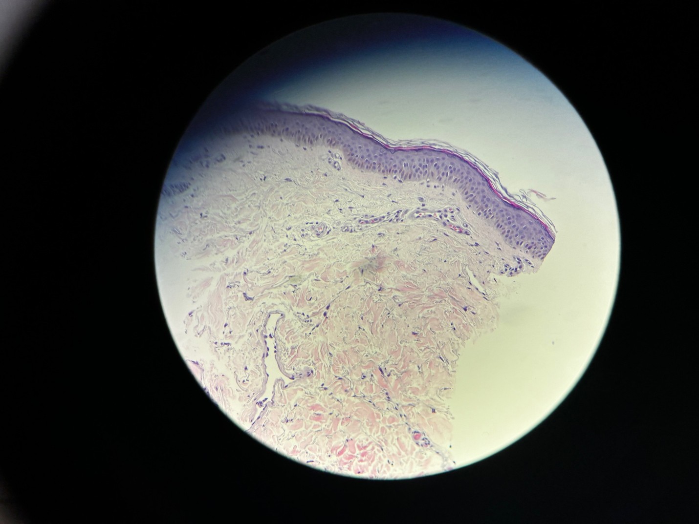
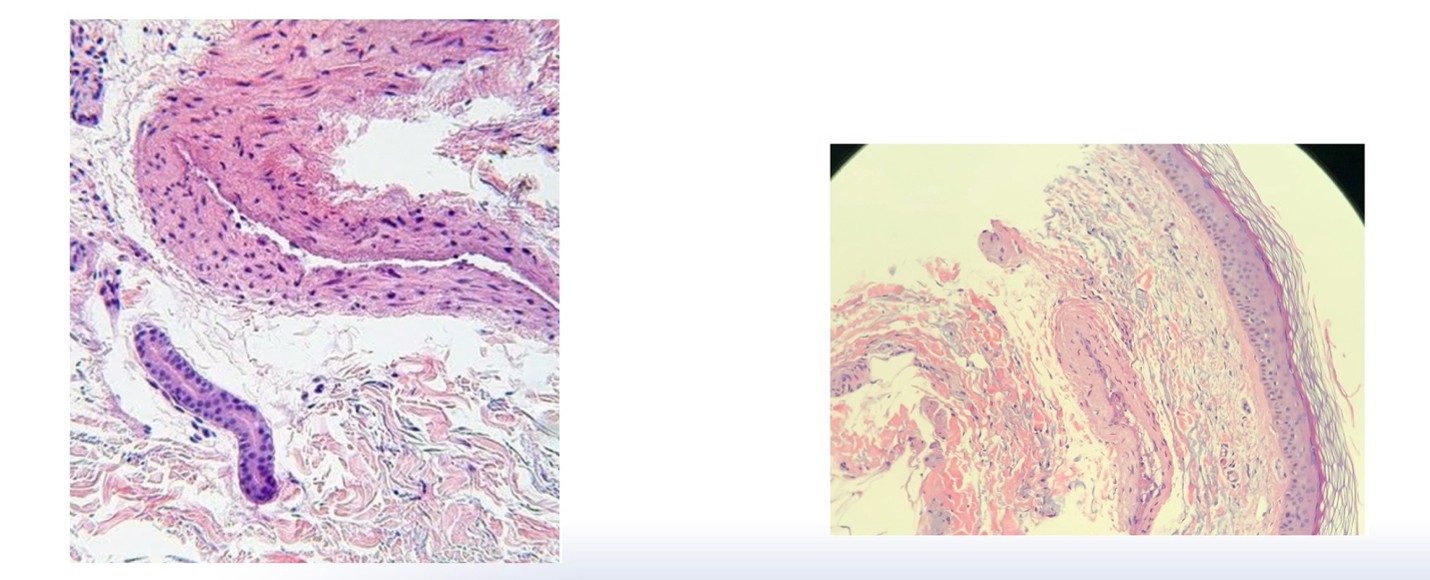
Before treating telangiectasias of the skin, it is imperative to understand the anatomy of the skin, particularly the dermis and its associated vessels.The region where most of the pathology occurs is the reticular dermis and the subpapillary plexus. The venous drainage of the reticular dermis is polygonal in distribution and rarely connects to adjacent regions, except in the case of aneurysmal telangiectasias.
Dermal veins lie approximately 500–600 microns below the skin surface and connect to ascending branches, also known as dermal perforators. When cutaneous venous hypertension develops, these veins dilate and their vessel walls thicken, often reaching approximately 500 microns in diameter. These dilated vessels are commonly recognized clinically as blue telangiectasias. The treatment of choice for blue telangiectasias is sclerotherapy using either liquid or foam sclerosants.
From a potency standpoint, Sotradecol® 0.1% diluted with saline is approximately equivalent to polidocanol 0.33%. However, Sotradecol® 0.1% diluted with bacteriostatic water to ½ normal saline (CMC) is approximately three times more potent than polidocanol 0.33%, making it a useful option for selected blue telangiectasias.
Red telangiectasias originate within or just below the subpapillary plexus. They may or may not be associated with underlying venous disease. When venous hypertension is the underlying etiology, vessel wall hypertrophy is often present. Unlike veins in the reticular dermis, pressure transmission may occur in either a horizontal or vertical direction. In generalized essential telangiectasia (GET), vessel walls may become hyalinized.
Treatment of red telangiectasias depends on their size, location, and underlying etiology. Available treatment modalities include vascular lasers, intense pulsed light (IPL), sclerotherapy, and microneedling.
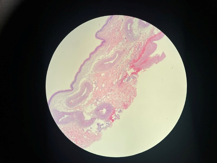
Reticular veins lie within the subdermal space approximately 2–3 mm below the skin surface. These veins transmit pressure to the dermis through ascending branches, or dermal perforators, and are often responsible for the cutaneous venous hypertension that contributes to the development of telangiectasias. When reticular veins are identified as the underlying source of venous hypertension, they are typically treated at the same session as the overlying spider veins. The preferred treatment is foam sclerotherapy, often combined with limited microphlebectomy when appropriate.
Understanding the origin and anatomy of fine veins is essential for selecting the most effective treatment. Matching the treatment modality to the specific type of telangiectasia improves outcomes and helps achieve optimal cosmetic results.